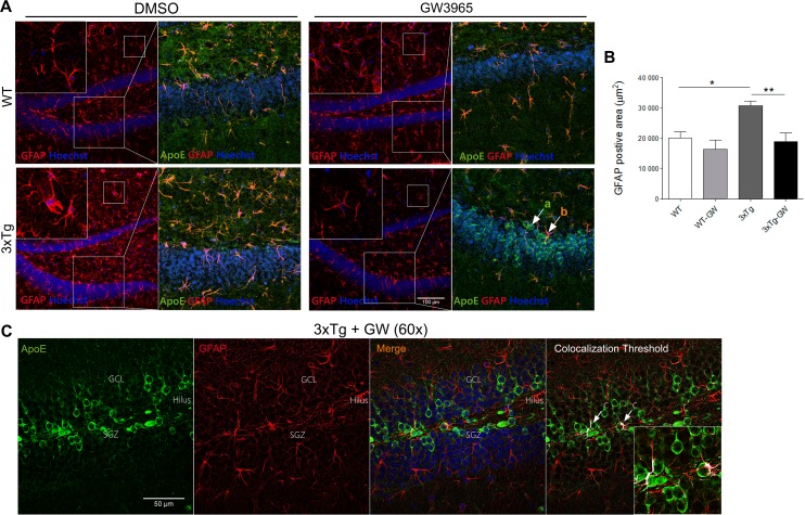
Fig 6. LXR activation reduces astrogliosis in treated 3xTg-AD.
(A) Representative microphotographs of GFAP (Red) immunohistochemistry and Hoechst and caption of granular layer showing ApoE (Green), GFAP (Red) and Hoechst (Blue) using confocal microscopy in the DG of hippocampal slices. Shown are: in the left upper panel WT mock (DMSO) treated slice showing baseline immunoreactivity. Right upper panel slice treated with GW, demonstrating no significant changes upon treatment. Lower panels are 3xTg-AD mice mock and GW treated slices, indicating a significant reduction of GFAP immunoreactivity upon GW treatment. Two different populations of cells are identified: (a) cells expressing ApoE and (b) cells expressing ApoE and GFAP. Also shown are close up views of molecular layer regions, which are included in the right upper corner of all images, to appreciate the change in morphology of astrocytes in 3xTg-AD treated with GW3965. (B) Quantitative analysis of the data presented in A, indicating the significant changes in GFAP positive areas produce by GW3965 in treated 3xTg-AD. Note that 3xTg-AD mice have a significant increased GFAP area, indicating astrogliosis, which is at baseline level after GW treatment. Statistical analysis was performed by one-way ANOVA followed by Bonferroni post test. (C) Representative close-up views (60X) micrographs of GFAP (Red) and ApoE (Green) immunofluorescence and Hoechst (Blue) of DG granular layer of treated 3xTg-AD mice under confocal microscopy. The colocalization threshold indicated in white showed a minimal population of cells with increased ApoE expression. Data are expressed as mean ± S.E.M. Differences are against untreated WT: *: p<0.05, **: p<0.01; n = 4 per group.

