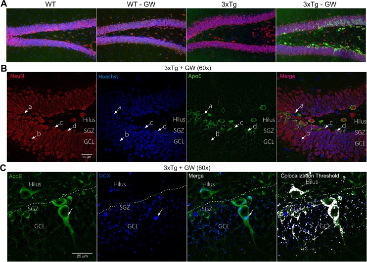
Fig 7. Neuronal localization of up-regulated ApoE in treated 3xTg-AD mice.
(A) Representative micrographs of DG showing the immunofluorescence of ApoE (Green), NeuN (Red) and Hoechst (Blue) of treated and untreated WT and 3xTg-AD mice using confocal microscopy (20X). (B) Representative micrographs of GL of DG of treated 3xTg-AD mice showing immunofluorescence of ApoE (Green), NeuN (Red) and Hoechst (Blue) at 60X magnification (C)) Representative micrographs of GL of DG of treated 3xTg-AD mice showing immunofluorescence of ApoE and Doublecortin (DCX) (Blue) at 60X magnification. n = 4 per group.

