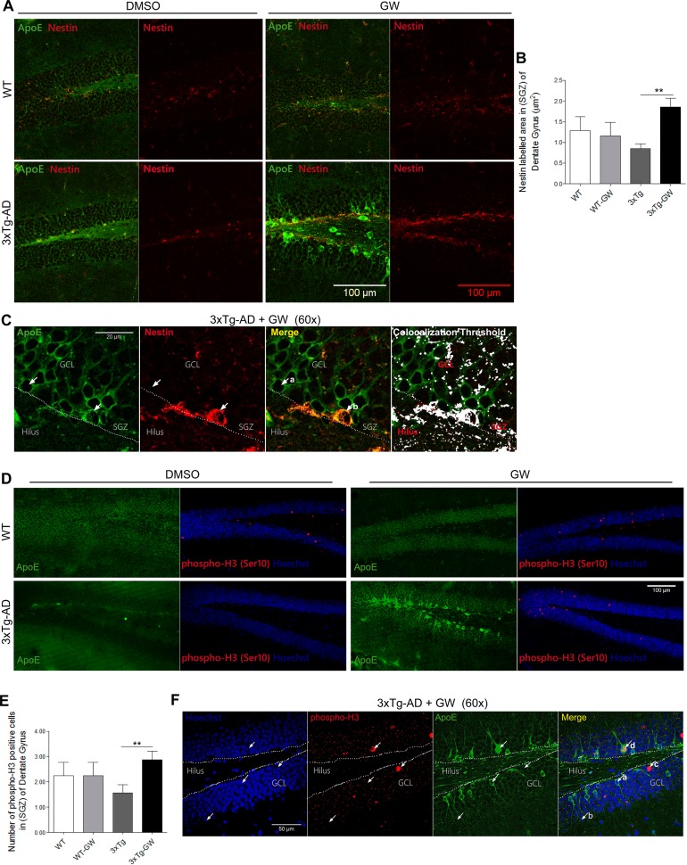
Fig 8. GW3965 increases the nestin immunoreactivity and the proliferating cells number in hippocampus of 3xTg-AD mice.
(A) Representative micrographs of multiple immunofluorescence of ApoE (Green) plus nestin (Red) and side by side only nestin (Red) immunofluorescence in DG using confocal microscopy for treated and untreated WT and 3xTg-AD mice. (B) Quantification of nestin positive immunofluorescence in the SGZ of DG. (C) Representative micrographs of ApoE (Green) and nestin (Red) immunofluorescence (60X), the colocalization threshold of ApoE and nestin indicates that there is a population of cells with increased ApoE expression. (D) Representative micrographs of DG of treated and untreated WT and 3xTg-AD mice showing the immunofluorescence of ApoE and side by side phospho-Histone H3 (Ser10) (Red) plus Hoechst (Blue). (E) Quantification of the number of positive-nuclei labeled with pHH3 (Red) and Hoechst (Blue). (F) Representative micrographs of ApoE (Green) and phospho-H3 (Red) immunofluorescence and nuclei Hoechst (Blue,60X), showing that there are proliferating cells with increased ApoE expression. Data are expressed as mean ± S.E.M. Statistical analysis was performed by one-way ANOVA followed by Bonferroni post test: *:p<0.05; **: p<0.01, n = 4 per group.

