Figure 1.
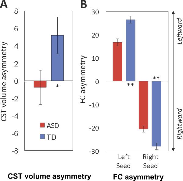
A. Leftward asymmetry of corticospinal tract (CST) volume observed in the typically developing (TD) group is significantly reduced in the group with autism spectrum disorders (ASD). Note: Bar graphs show marginal means ±1 standard error of the mean after covarying for age, total motion index, and total brain volume. B. Both the left and right precentral gyrus seeds show significant reductions in asymmetry of functional connectivity in the ASD compared to the TD group. In both panels, leftward asymmetry is up (positive values on y axis); rightward asymmetry is down (negative values). *p<.05; **p<.001.

