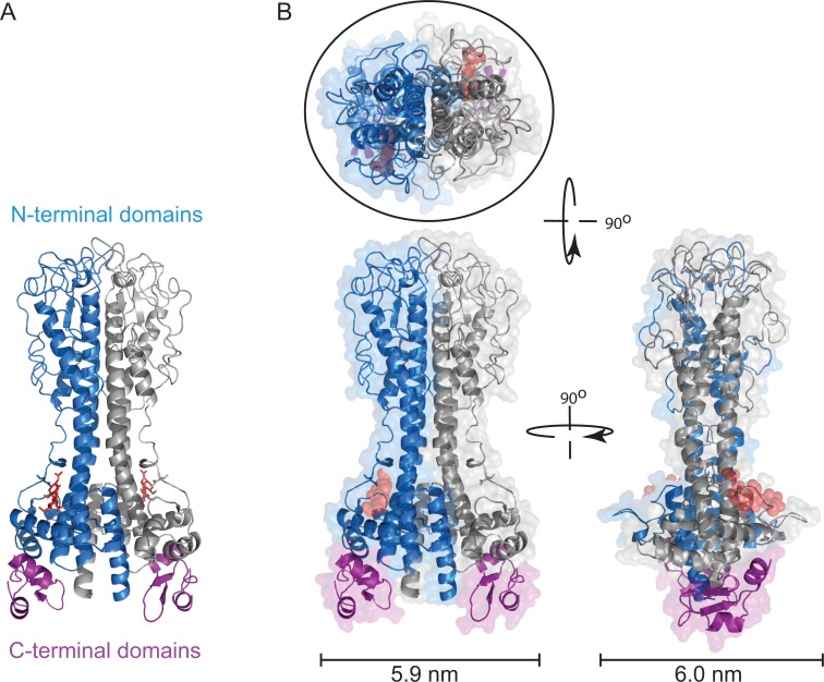
Fig 1. Structure of VSG221 (MITat1.2).
(A) Illustrated model of VSG221 dimer showing the structures of the N-terminal domain, one monomer in blue and one in grey, and the two C-terminal domains in purple (PDB: 1VSG and 1XU6) [1,9]. The N-linked oligosaccharide in the N-terminal domain is shown in red. Three residues are shown that form the core; there are between one and three further residues not shown. The relative positions of the N- and C-terminal domains are not known, and this illustration is a model [1]. (B) Space-filling model of VSG221 viewed from the x-, y-, and z-axes. The maximum width dimensions of the VSG are shown below, and the fit of an ellipse of approximately 28 Å2 is shown around the structure of a VSG viewed from outside the cell.

