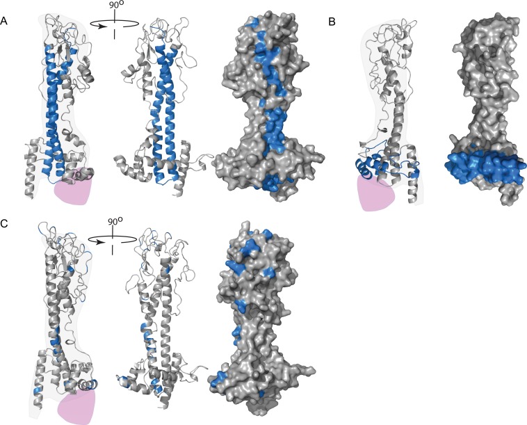
Fig 3. VSG models.
(A) A model of VSG121 showing the location of the cyanogen bromide fragment p19 (blue) that contains the epitopes for MoAbs that bound live trypanosomes. From the left, one monomer orientated so the dimerization interface runs vertically up the page; second, rotated approximately 90° so that the dimerization interface has turned away from the observer; third, same view with the surface added. There are potential surface-exposed epitopes along the entire length of the domain. (B) A model of VSG117 showing in blue the location that contained the epitope recognised by a MoAb that bound live cells. (C) Model of VSG WATat1.1 showing the location of differences with the related VSG WATat1.12. A monoclonal antibody that recognises an epitope in WATat1.1 does not bind WATat1.12, so the epitope probably contains one of these differences. An envelope for one possible position of the C-terminal domain is shown in purple.

