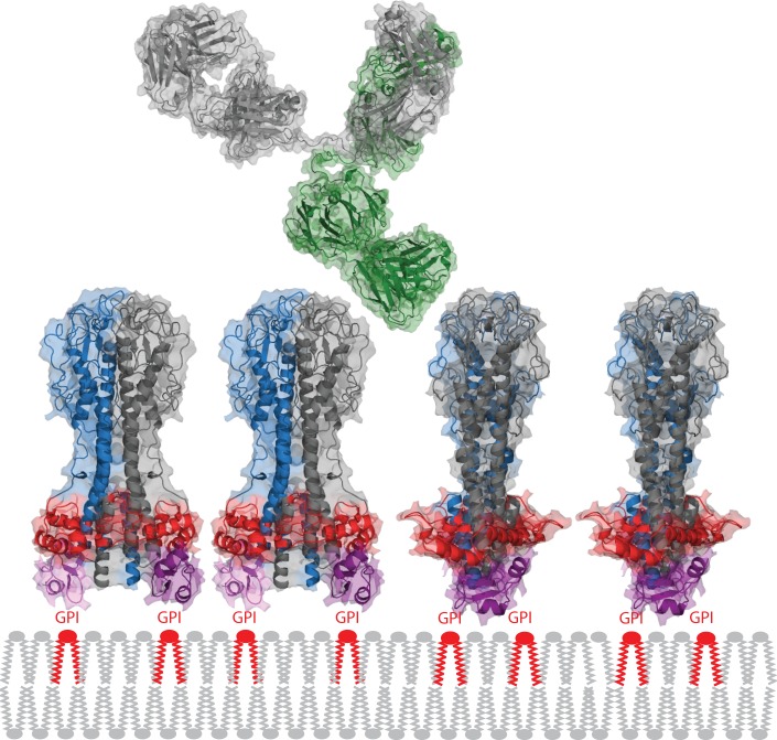
Fig 4. Packing of the VSG on the plasma membrane and comparison with an immunoglobulin G.
The VSG spacing is based on experimentally determined copy number and surface area estimates. The widest part of the VSG N-terminal domain is shown in red and the C-terminal domain in purple. Together, these two features probably form the barrier that restricts access of immunoglobulins to the plasma membrane. One heavy and one light chain in the IgG is shown in green, the other pair in grey. The VSG structure is derived from PDB 1VSG and 1XU6, the IgG from PDB 1IGY.

