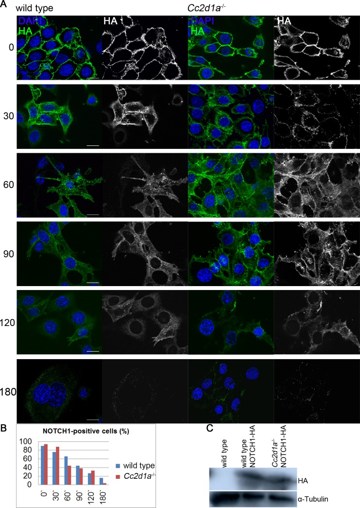
Fig 6. NOTCH1 endocytosis and degradation in wild type and Cc2d1a deficient MEFs.
(A) Pulse-chase antibody uptake assay. NOTCH1-HA overexpressing MEFs were treated with Alexa 488 coupled anti-HA antibody and endocytosis and degradation was monitored for various points in time. (B) Quantification of NOTCH1 degradation. Stated is the average percentage of cells showing HA staining after the analysed incubation times. A minimum of 50 cells per point in time from at least 2 independent experiments were counted. (C) Immunoblotting of protein lysates from indicated MEF cells. The 300kDa NOTCH1-HA band is detected in transduced cells, but not in wild type cells. Scale bars are 20 μm.

