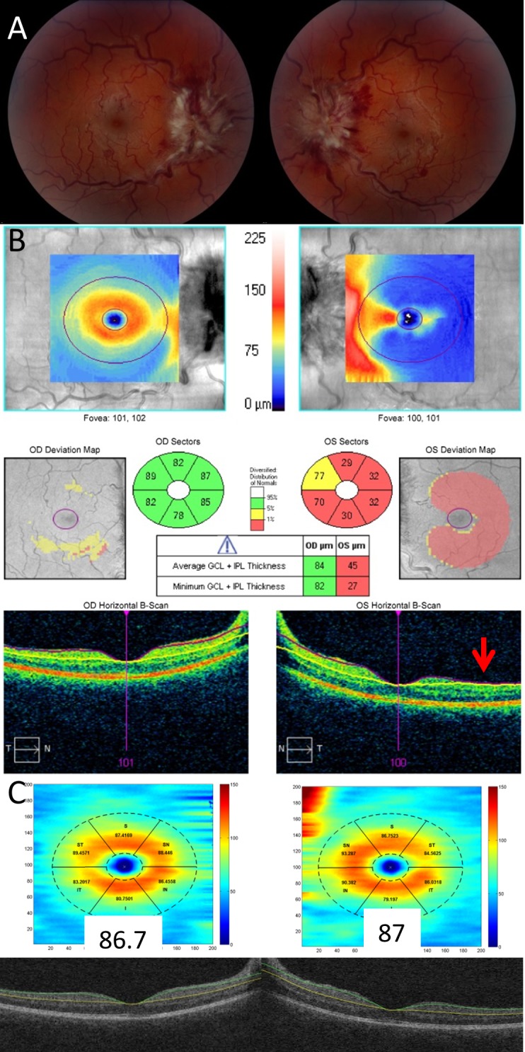
Figure 1.

Comparison of the Cirrus and Iowa segmentation algorithms of the GCL-IPL complex in a patient with significant optic disc edema. Fundus photography demonstrates grade IV optic disc edema in both eyes (A). Cirrus segmentation of the GCL-IPL complex correctly segments the right eye, but fails to correctly segment the left eye where the segmentation lines collapse upon one another (red arrow) giving rise to artifactual GCL-IPL thinning (B). The Iowa Reference Algorithm correctly segments the GCL-IPL complex in both eyes (C).
