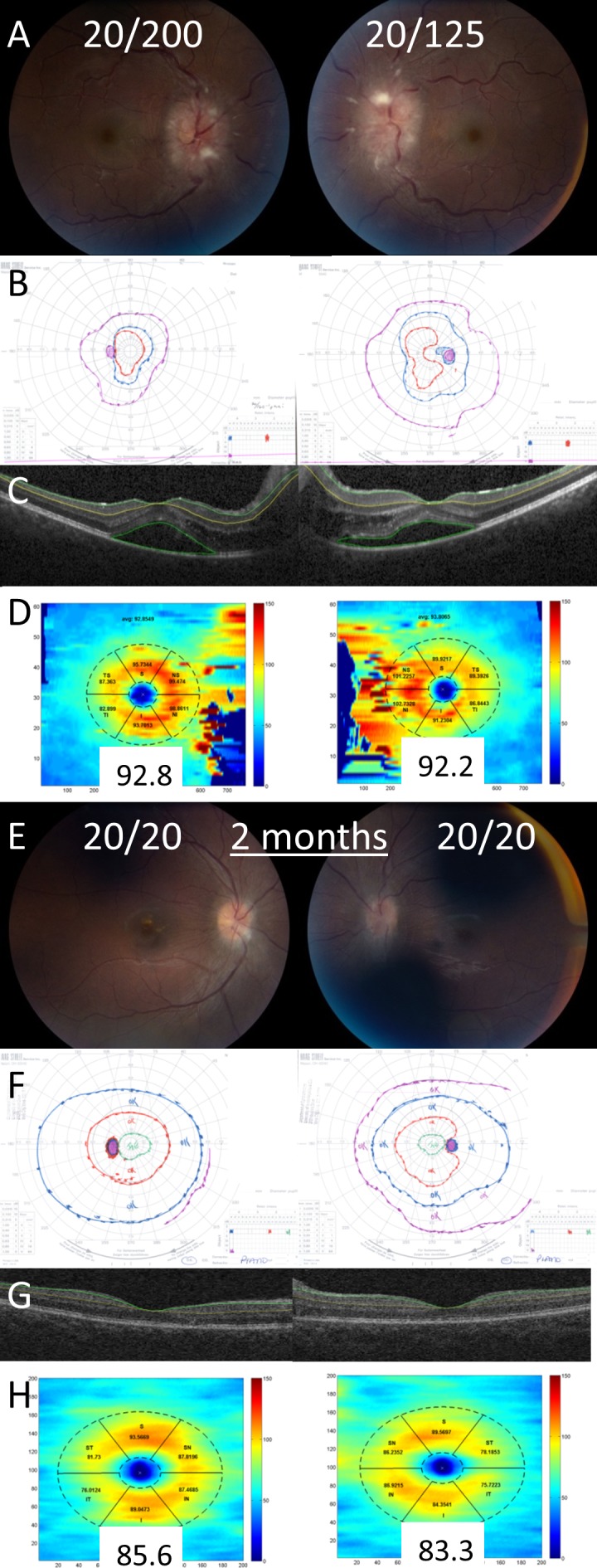
Figure 4.

Example of a patient with decreased visual acuity from subretinal fluid. On presentation, the patient had visual acuities of 20/200 in the right eye and 20/125 in the left eye, grade IV optic disc edema in both eyes (A), and constricted Goldmann visual fields (B). Optical coherence tomography of the macula demonstrates subretinal fluid in both eyes (C). The GCL-IPL complex was segmented with the Iowa Reference Algorithm (C) and the average thickness was normal in both eyes (D). After 2 months, visual acuity improved to 20/20 in both eyes, the optic disc edema was significantly improved (E), and the Goldmann visual fields were normal (F). The GCL-IPL was segmented using the Iowa Reference Algorithm (G) and the average thickness remained normal in both eyes (H).
