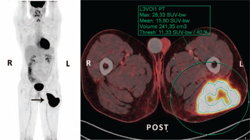
FIGURE 1.

36-year-old male with high-grade soft tissue sarcoma (subclassification not possible; tumor grade III [FNCLCC grading system]) located in the posterior musculature on the left femur (arrow). The patient underwent a preoperative F-18 FDG PET/CT scan for staging, which showed no metastatic disease. For volume-based calculations of tumor metabolic activity, a volume-of-interest (VOI) was manually drawn on the acquired PET images (green circle), and estimation of the maximum standardized uptake value of primary tumor was performed (SUVmax 28.33 g/mL). Dedicated software and a preset threshold of 40% of SUVmax of primary tumor (SUV 11.33 g/mL) was used to define the metabolic tumor volume (irregular green shape; MTV40% 241.35 mL), and the mean standardized uptake value of the MTV40% was determined (SUVmean 15.80 g/mL). The 2 latter variables were used for calculation of total lesion glycolysis (TLG = MTV40% × [SUVmean of the MTV40%] = 3813.33 g). A wide resection of tumor (12 × 7 × 10 cm) was achieved. No adjuvant therapy was administered. One year after surgery the patient presented with metastatic disease and he died 2 months later. CT = computed tomography, F-18 = fluorine-18, FDG = fluoro-2-deoxy-D-glucose, FNCLCC = French Federation of Cancer Centers Sarcoma Group, MTV = metabolic tumor volume, PET = positron emission tomography, SUVmax = maximum standardized uptake value, TLG = total lesion glycolysis, VOI = volume of interest.
