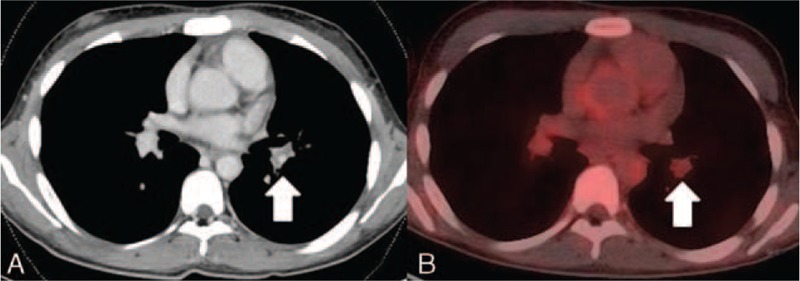
FIGURE 1.

(A) Defect of contrast in the pulmonary artery without narrowing of the pulmonary artery, which suggested thrombosis (arrow). (B) PET showed no FDG accumulation in the wall of the same lesion of the pulmonary artery (arrow). FDG = 18F- fluoro-2-deoxy-D-glucose, PET = positron emission tomography.
