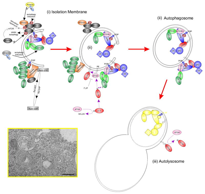
Fig. 1.
Simplified schematic diagram of autophagy with key molecular machinery. Autophagy is a constitutive catabolic mechanism responsible for the turnover of all cellular constituents. With stress (such as starvation), autophagic flux is accelerated. A double membrane ‘isolation membrane’ (ii) forms and fuses as an ‘autophagosome’ (ii) to capture damaged proteins, organelles and cytoplasm. Degradation follows fusion with a lysosome as an ‘autolysosome’ (iii; Figure expanded from Fig. 3B of Wang et al, ‘13). Inset, electron micrography of forming autophagosome-like structures in human corneal epithelial cells stressed with INFG and TNF in the presence of transient autophagic stimulator lacritin, Bar = 0.5 μM.

