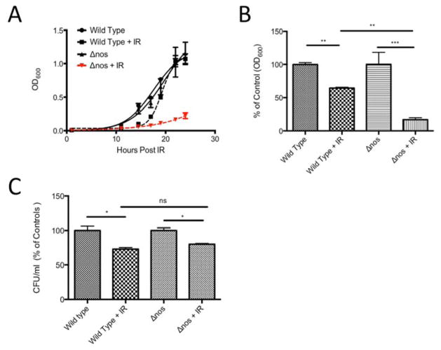
Figure 1. Δnos is more susceptible to ionizing radiation than wild type Drad.
Panel A: Growth curves (monitored by OD600) are depicted. Dashed lines indicate growth of wild type nos and Δnos Drad following exposure to extreme ionizing radiation (γ-radiation, 3000 Gy). Cultures were grown to OD 0.8–1.0 and irradiated followed by 1:100 dilution in fresh TGY media and monitoring of growth. Panel B: Relative growth of Δnos and wild type nos Drad with and without 18hrs post-irradiation. Panel C: The percentage of viable cells in post-irradiated samples from Panel B was determined based on colony forming assays (CFU/ml) and compared to controls. Data are plotted as mean +/− SD of three biological replicates. Statistical significance was assessed using one-way ANOVA and Tukey post-hoc testing. (*p<0.05, **p<0.01, ***p<0.001).

