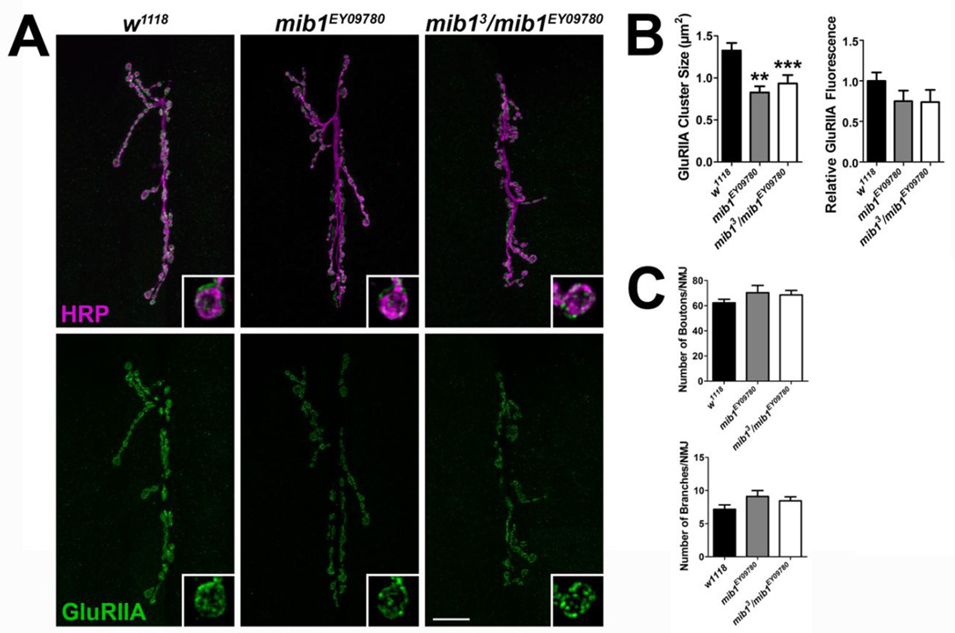
Figure 3. Mib1 is important for the clustering of GluRIIA-containing receptors.
(A) Control and mib1 mutant confocal images showing representative 6/7 NMJs from third instar larvae. Preparations were immunolabled with α-HRP to label presynaptic motor neurons (magenta) and α-GluRIIA (green) to label GluRIIA-containing glutamate receptor clusters. Inset panels show high resolution terminal boutons. Scale bar = 20 µm. (B) Histograms showing quantification of GluRIIA cluster sizes (left) and GluRIIA relative fluorescence intensity (right) for genotypes shown in A. (C) Quantification of characteristics representative of presynaptic motor neuron morphology including the number of boutons (top) and branches (bottom).

