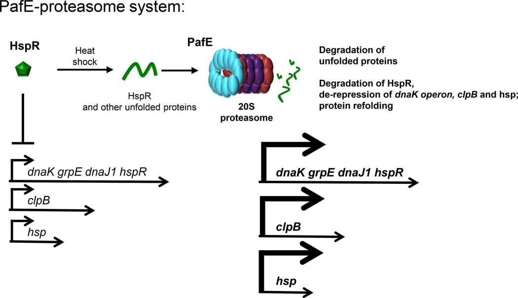
Figure 2. The PafE-proteasome System of M. tuberculosis.
PafE (blue) forms dodecameric rings and caps 20S CPs. This allows the ATP-independent opening of 20S CPs to facilitate the degradation of peptides and denatured proteins as well as HspR. HspR represses expression of the dnaK operon, clpB and hsp (Rv0249c). See text for details.

