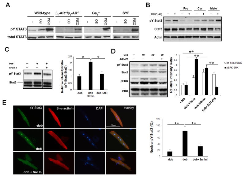
Figure 2.
Activation of STAT3 by βARs requires G αs and Src-family kinases. A, Western blot analysis of STAT3 activation in response to ISO (1 μM, 30 minutes) in β1AR/β2AR-compound deficient, G αs-deficient, and Src family kinases (SYF)-deficient mouse embryonic fibroblast (MEF) cells. OSM treatments were used as a positive control for pY-STAT3. B, Western blot analysis of STAT3 activation in neonatal cardiomyocytes in response to ISO (1 μM, 30 minutes) that pre-incubated with different βARs antagonists for 1 hour. Pro, propranolol; Car, carvedilol; Meto, metoprolol. C, Western blot analysis of pY-STAT3 in mouse neonatal cardiomyocytes with or without pre-incubation of Src kinase inhibitor I (1 μM) for 15 minutes, followed by dobutamine (1 μM) treatment. The relative intensity ratios between pY-STAT3 and total STAT3 are shown in the right panel, *P<0.05. D, Western blot analysis of pY-STAT3 and pERK activation in mouse neonatal cardiomyocytes with or without pre-incubation of EGFR inhibitor AG1478 (1 μM) followed by dobutamine (1 μM) treatment. The relative intensity ratios of pY-STAT3 vs total STAT3 and pERK1/2 vs total ERK1/2 are shown in right panel, **P<0.01 E, Representative fluorescence images of immune reactivity to anti-pY-STAT3 antibody (in green) in isolated wild-type cardiomyocytes treated with dobutamine (1 μM) for 30 minutes with or without pre-incubation of Src kinase inhibitor I (1 μM). α-actinin, in red; DAPI, in blue. The experiments were independently repeated 3 times (N >300 cells/each group) and results are expressed as positive cell number over total cell number (Right panel), **P<0.01.

