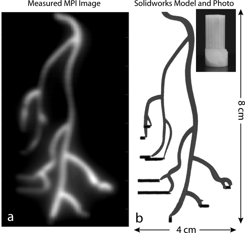
FIG. 1.
As a tracer imaging technique, MPI has applications in molecular imaging and angiography. (a) Experimental MPI image of (b) 3D printed coronary artery model. The modeled arteries (1.8–2.3 mm diameter) formed cavities within the cylindrical 3D ABS plastic model with injection holes illustrated in black and are filled with one part SPIO tracer (Nanomag-MIP) and four parts DI water. The maximum intensity projection image was acquired in the Berkeley 3D MPI scanner with a 10 min total imaging time and a 4.5 × 3.5 × 9.5 cm field-of-view. No deconvolution was performed. A threshold at 10% of the maximum signal was applied to remove noise.

