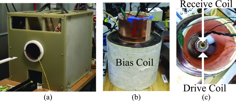
FIG. 3.
(a) The Berkeley 3D MPI scanner acquires 3D images using a 7 T/m selection field. A drive coil scans the FFP of the scanner at 23.2 kHz up to 30 mT peak amplitude. The Berkeley relaxometer, shown with (b) side and (c) top views, measures the PSFs of SPIO nanoparticles. A sinusoidal magnetic field is generated in the drive coil at frequencies of 1.5–25 kHz and of 5–100 mT drive field peak amplitude, and a bias coil of ±160 mT. The MPI signal is detected using a gradiometrically decoupled receive coil and digitized at 10 MSPS.

