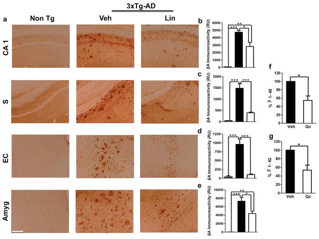
Figure 4. 3xTg-AD linalool-treated mice show reduced β-amyloidosis.
a) Representative images of βA (anti-βA 6E10) immunoreactivity in the CA1 area, the subiculum, the EC and the amygdala of vehicle- and linalool-treated 3xTg-AD and vehicle-treated Non-Tg mice at 21–24 months of age. Magnification: 10x; scale bar: 50 μm. b) The values in the bar graph are expressed in densitometric relative units (RU) of βA immunoreactivity in the CA1 area, c) he subiculum, d) the EC and e) the amygdala. f) The relative βA 1–40 and g) βA 1–42 fragment levels were analyzed in hippocampal lysates by ELISA. Veh: vehicle (Saline solution); Lin: Linalool; CA1 and S: subiculum of the hippocampus; EC: entorhinal cortex; Amyg: amygdala. The data are expressed as the means ± SEM. n=4–5. *p<0.05; **p<0.01; ***p<0.001.

