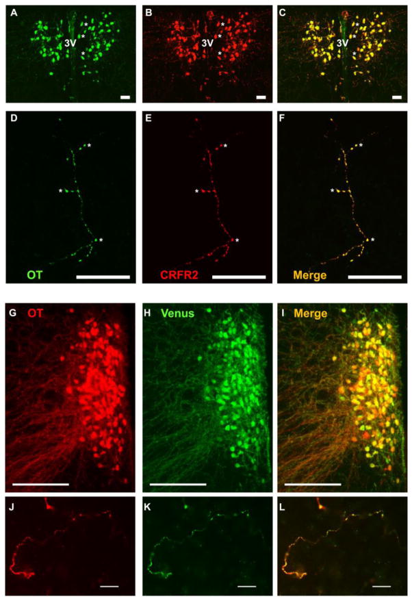
Figure 3.
Co-localization of OT and CRFR2 (A – F) or Venus (G – L) in the prairie vole PVN and NAc shell.
The PVN exhibits high somatodendritic immunoreactivity of OT (A; green) and CRFR2 (B; red) in neurons flanking the 3rd ventricle (3V). Asterisks (*) in A – C mark cells within the right hemisphere that clearly reveal overlapping (C; yellow), but nonidentical, immunoreactivity. This pattern appears equivalent to the double-labelling described in detail in rats (Dabrowska et al., 2011). Sparse large-caliber neuronal fibers that extend into the NAc shell also exhibit strong co-localization of OT (D) and CRFR2 (E; overlap F). Asterisks (*) in D – F mark distinct co-labeled puncta.
In the PVN, both OT (G; red) and Venus (H; green) immunoreactivity were co-localized in one hemisphere of the PVN exhibiting a robust plume of OT neurons (merge: I; yellow). In the NAc shell, both OT (J; red) and Venus (K; green) immunoreactivity were co-localized (merge: L; yellow).
Scale bars = 20 μm (A–F; J – L) and 200 μm (G – I)

