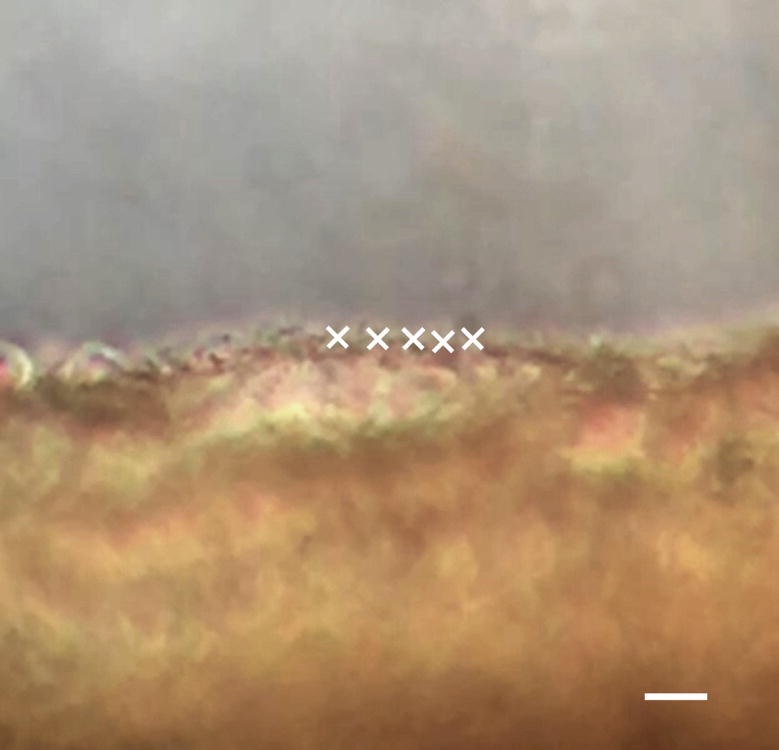
Figure 2. CBF analysis using phase-contrast microscopy at 32× magnification.

This is a cropped image segmented from a high speed digital video recorded at 240 fps using phase-contrast microscopy. The MATLAB program developed in the lab allows the user to manually select points of interest along the ciliated epithelium, as indicated by the white X’s. The scale bar indicates 10 μm.
