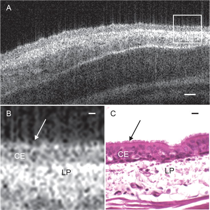
Figure 4. Functional anatomy of rabbit tracheal mucosa.

(A) Cropped OCT B-scan captures a 2D-crossectional image of ex vivo rabbit tracheal mucosal tissue at approximately 6 μm axial resolution and 25 μm lateral resolution. (B) Magnified region outlined in white box of tracheal mucosa in figure 4(A) to show tissue substructure. (C) Corresponding histology of rabbit tracheal mucosa. Arrows indicate the surface of the epithelial layer where the cilia reside. CE = ciliated epithelium, LP = lamina propria. Scale bar in (A) indicates 50 μm. Scale bars in (B) and (C) indicate 10 μm
