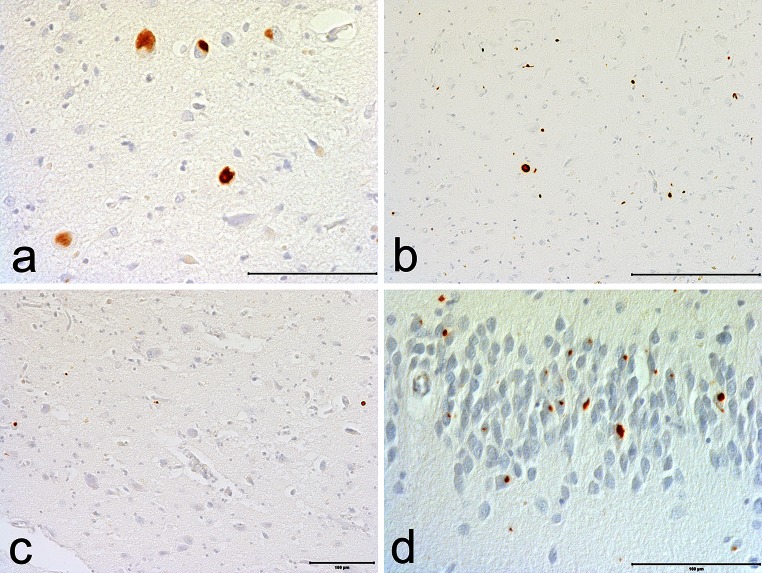
Fig. 4.
pTDP-43 pathology in CTE. a pTDP-43 neuronal inclusions in the amygdala. b p-TDP-43 inclusions and dot-like neurites in CA1. c p-TDP-43 dot-like neurites in entorhinal cortex. d pTDP-43 inclusions and dot-like neurites in the dentate granule cell layer. All sections immunostained for p-TDP-43, bars indicate 100 µm

