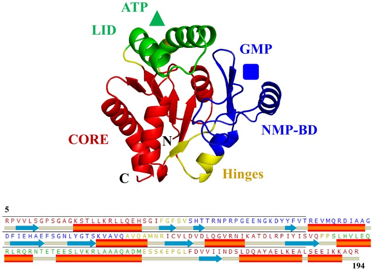
Fig. 1.
The Swiss homology model for hGMPK was constructed by using the crystal structure of mGMPK’s closed conformation (residues 5-194 of 197 aa) as template [pdb 1LVG (Sekulic et al. 2002)]. The three major structural regions present in hGMPK are designated and color-coded as the NMP-binding domain (NMP-BD) (blue), the CORE (red), and the LID (green). These structural regions in hGMPK (residues 5-194 of 197 aa) are interconnected by four hinges (yellow). hGMPK substrates, GMP (blue square), which bind to NMP-BD and substrate ATP (green triangle), which bind to LID in GMPK are schematically presented

