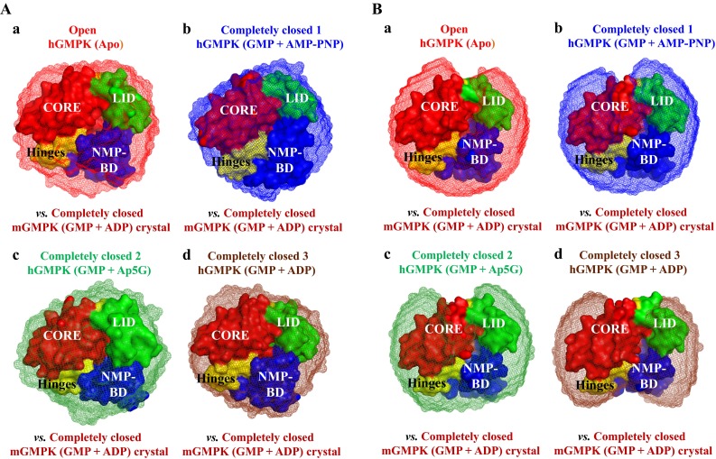
Fig. 4.

Overlay of reconstructed hGMPK SAXS models (mesh representation) on the crystal structure of the completely closed mGMPK conformation (GMP + ADP, pdb 1LVG (Sekulic et al. 2002), surface representation). GASBOR (A) and DAMMIN (B) SAXS models of different hGMPK conformations are compared separately. a Open hGMPK conformation (apo-form). b Completely closed hGMPK conformation 1 (GMP + AMP-PNP). c Completely closed hGMPK conformation 2 (Ap5G). d Completely closed hGMPK conformation 3 (GMP + ADP). Overlay was done with SUPCOMB13 and the resulting NSD values are shown in Table S4, ESM. Different structural regions in the crystal structure of completely closed mGMPK (NMP-BD, CORE, and LID) and hinges are color-coded
