Fig. 1.

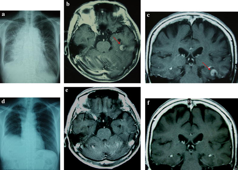
Chest X-ray examination and T1-weighted magnetic resonance imaging (MRI) with gadolinium. a Chest X-ray examination shows consolidation of the lung and pleural fluid. b and c Axial and coronal T1-weighted MRI with gadolinium. An enhanced mass lesion (red arrows) in the left temporal lobe was observed on admission. d The consolidation of the lung and pleural fluid was not detected 1 month after the administration of gefitinib. e and f Brain metastasis was not detected 1 month after the administration of gefitinib
