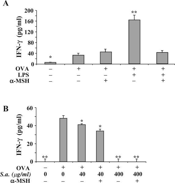
Fig. 2.
The effects of α-MSH on antigen-specific APC presentation under the influence of specific TLR-stimulation. Adherent spleen cells were treated with α-MSH (30 pg/ml) and pulsed with OVA (200 μg/ml) that was mixed with (A) LPS (1 μg/ml) or (B) S. aureus (200 μg/ml). The cells were washed and OVA-antigen primed T cells (4×10⁵ cells/well) were added to the cultures. The amount of IFN-γ produce in the cultures 48 h later was assayed by ELISA. The results are presented as the mean pg/ml±S.E.M. of IFN-γ measured in the cultures of 4 independent experiments. Significant differences (*P<0.001, **P<0.0001) were calculated in comparison with the IFN-γ concentration in cultures of T cells activated with APC pulsed with uncontaminated OVA and not treated with α-MSH. The difference in panels (A) and (B) between the IFN-γ concentrations in cultures of T cells activated with APC pulsed with uncontaminated OVA and not treated with α-MSH is insignificant.

