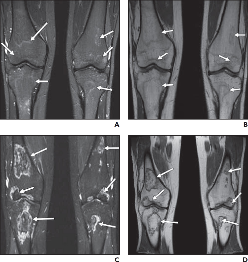
Fig. 1. 17-year-old boy.
A and B, On STIR (A) and T1-weighted (B) coronal images of knees, signal changes (arrows) are seen in distal femoral epiphyses (> 30% articular involvement), proximal tibial epiphyses, and metaphyses bilaterally.
C and D, STIR (C) and T1-weighted (D) coronal images of knees at 3-month follow-up examination show evolution of previous signals into typical osteonecrotic lesions (arrows) conforming to original signal pattern.

