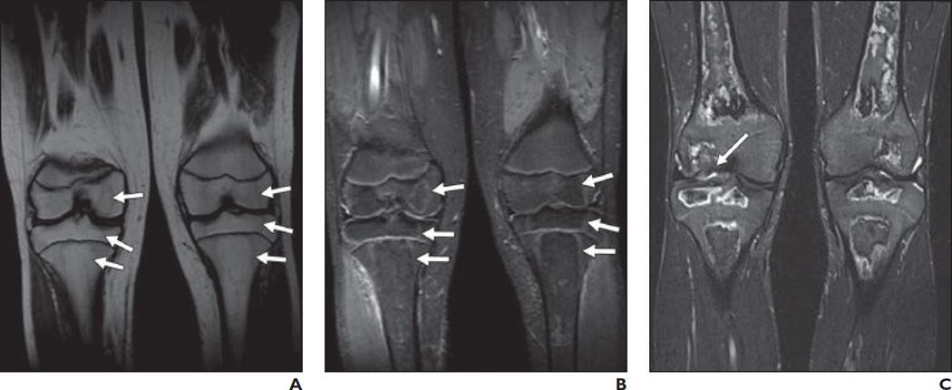
Fig. 4. 14-year-old boy.
A and B, On T1-weighted (A) and STIR (B) coronal images of knees, signal changes (arrows) involving > 30% epiphyseal articular surfaces of tibial and femoral condyles are seen bilaterally.
C, STIR coronal knee image on follow-up MRI performed 6 years later shows articular cartilage irregularity (arrow) involving right femoral condyle.

