Fig. 2.
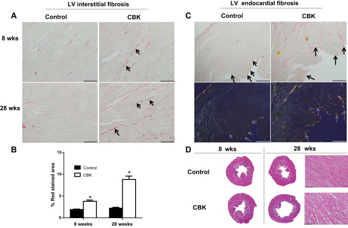
CBK mice triggered collagen deposition and dilation in left ventricle (LV) compared with controls. A: LV middle region images (40× magnification) stained with picrosirius red (PSR) at 8 and 28 wk for wild-type (WT) and CBK mice indicated higher interstitial collagen deposition in CBK mice at 28 wk. Intense red color indicates collagen deposition. B: quantification of collagen density in CBK mice compared with control mice at 28 wk of age; 4–5 images/mouse, n = 5 mice/group. C: 10× magnification images of PSR-stained 28 wk WT and CBK mice show interstitial and endocardial fibrosis (black arrows, top panel). Under plane-polarized light endocardial fibrosis is marked by an orange color (white arrows, bottom panel). D: hematoxylin- and eosin (H&E)-stained LV middle cavity (1.25× magnification) indicates hypertrophy and dilation at 28 wk compared with age-matched control. H&E LV staining indicates dilative hypertrophy at 28 wk of age (40× images). *P < 0.05 vs. age-matched controls (CON).

