
Fig. 4.
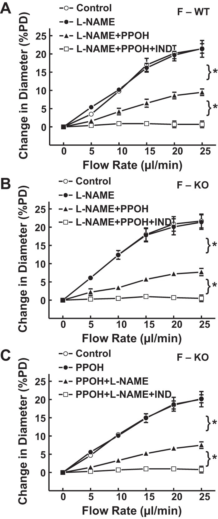
A: normalized diameter of gracilis muscle arterioles in F-WT mice (n = 6), as a function of perfusate flow in the control condition, in the presence of l-NAME, l-NAME + PPOH, and with additional IND. B and C: normalized diameter of gracilis muscle arterioles in F-KO (n = 8 and 9) as a function of perfusate flow, in the control condition, in the presence of l-NAME or PPOH alone, combination of the 2 inhibitors, and with additional IND mice. *Significant difference between the 2 curves.