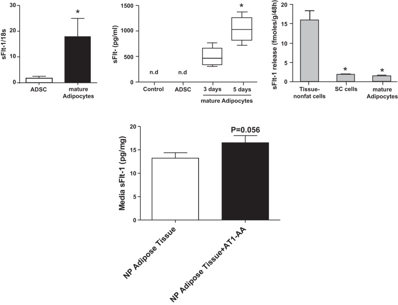
Fig. 6.
Top, left: mRNA expression of sFlt-1 in isolated adipose-derived stem cells (ADSCs) and isolated mature adipocytes from human adipose tissue; *P < 0.01. Middle: release of sFlt-1 from ADSCs incubated for 5 days and mature adipocytes incubated for 3 or 5 days; *P < 0.05 for 3 vs. 5 days; n.d., not detectable. Right: release of sFlt-1 from adipose tissue nonfat cells, stromal vascular cells (SV cells) and mature adipocytes; *P < 0.01 vs. adipose tissue nonfat cells. From Herse et al. (66). Bottom: stimulation with AT1-AA promotes sFlt-1 release from adipose tissue explants isolated from normal pregnant (NP) Sprague-Dawley rats. Adipose explant (∼100 mg) were incubated in F12K supplemented with media (DMEM containing 4 mM l-glutamin, 4,500 mg/l glucose, 1 mM sodium pyruvate, 1,500 mg/l sodium bicarbonate, 4 g/l BSA, and 1% PenStrep) in the presence or absence of AT1-AA (1:100 dilution) for 48 h, at which time media were collected and assayed for sFlt-1 as previously described (171). n = 3 rats/group. Data are means ± SE. Data in bottom panel are unpublished.

