Fig. 3.

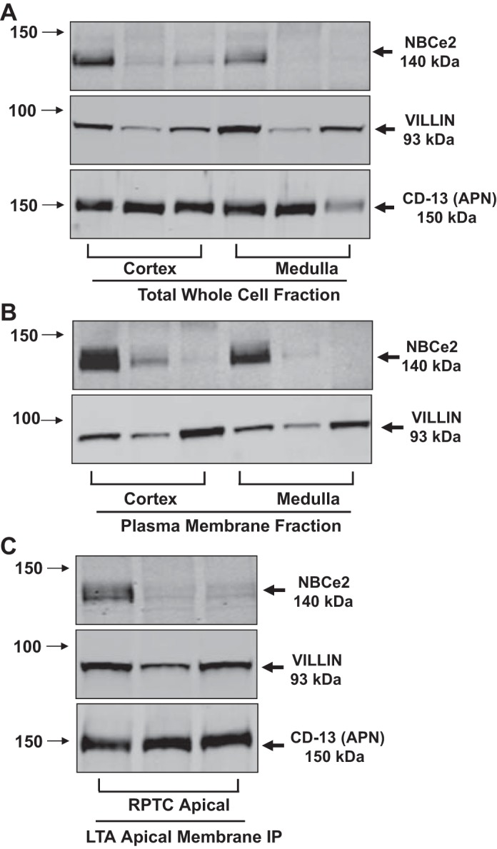
Western Blot analysis of total whole cell homogenates and membrane fractions from human kidney tissue. A–C: results from three separate patient samples. Arrows on the left side of the blots indicate the molecular sizes, while the arrows on the right side indicate the bands of the proteins of interest. Villin was used as a RPTC microvilli marker. CD-13 (APN) was also used in A and C as another RPTC-specific, brush-border membrane marker. A: Western blot analysis of total whole cell fractions from cortex and medulla. The blots were probed with NBCe2, villin, and CD-13 antibodies. B: Western blot analysis of plasma membrane fractions from the cortex and medulla: The blots were probed with NBCe2 and villin antibodies. C: Western blot analysis of LTA apical membrane fractions from cortex. The blots were probed with NBCe2, villin, and CD-13 antibodies. A clear signal in the Lotus tetragonobulus agglutinin (LTA)-immunoprecipitated (IP) apical membrane fraction, along with two different RPTC apical membrane markers, suggests that NBCe2 is expressed in the RPTC apical membrane, including brush border in human kidneys.
