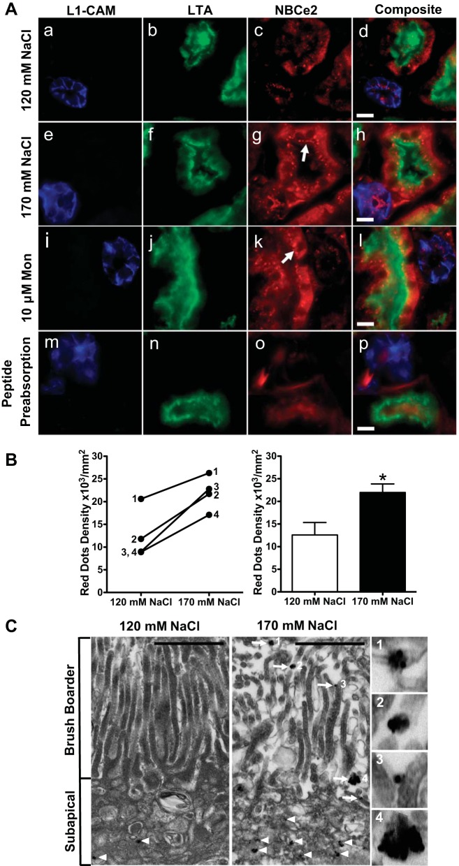
Fig. 5.
Change in localization of NBCe2 following an increase in extracellular sodium in kidney tissue slice culture. A: effect of an increase in extracellular sodium on NBCe2 localization. Fresh human renal cortical tissue slices (1-mm thickness) were cultured for 30 min in media containing low (120 mmol/l NaCl; top), or high (170 mmol/l NaCl, second row) extracellular sodium. They were also incubated for 1 h in media containing 10 mmol/l monensin (third row). Slices were fixed, and 8-mm frozen sections were made. The cortical collecting duct (CCD) marker, L1-CAM (blue; a, e, i, and m), the proximal tubule brush border marker, LTA lectin (green; b, f, g, and n), and NBCe2 antibody (red; c, g, k, and o) confirmed NBCe2 staining in both proximal tubule and CCD. In 120 mmol/l extracellular NaCl concentration, NBCe2 localization is predominantly cytoplasmic (C), while in increased (170 mmol/l) NaCl concentration or in the presence of monensin, NBCe2 is also localized in punctate subapical structures (g and k, white arrow), with some colocalizing with LTA (yellow orange). After peptide preadsorption (m–p), NBCe2 immunostaining is markedly decreased. The tissue for peptide preadsorption was incubated in 170 mmol/L NaCl. Scale bar = 10 μm. B: quantification of images from the low and high extracellular sodium experiments shown in A. Kidney slices from four separate patient samples were analyzed by counting the red dots from all the proximal tubules and measuring the area (dots/mm2). Changes following the increase in extracellular sodium from 120 to 170 mmol/l NaCl are shown for each individual patient on the left graph, and the summary is shown on the right graph. A significant increase in NBCe2 staining in the subapical area of proximal tubules (as identified by LTA) is shown (n = 4, *P < 0.05). C: immuno-electron micrograph of NBCe2 in human kidney cortex. Ex vivo incubation of fresh human kidney cortex under low or elevated NaCl concentrations produced an increase in brush border-localized immune-reactive electron dense puncta on the microvilli of the proximal tubule, indicated by arrows. C: enlarged photos in panels 1–4 to demonstrate that NBCe2-immunoreactive particles appear in clusters. A notable increase in puncta in the microvilli is found under elevated sodium conditions. Scale bar = 1 μm.

