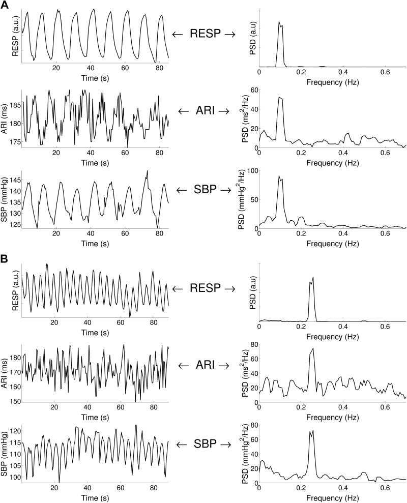
Fig. 3.
Respiratory-related ARI and SBP oscillations. A: example of ARI, SBP, and respiration (RESP) recordings for 0.1 Hz (6 breaths/min) breathing. Cyclical variation was observed in both ARI and SBP at the breathing frequency. PSD, power spectral density. B: example of ARI, SBP, and RESP recordings for 0.25 Hz (15 breaths/min) breathing. Cyclical variation was observed in both ARI and SBP at the breathing frequency.

