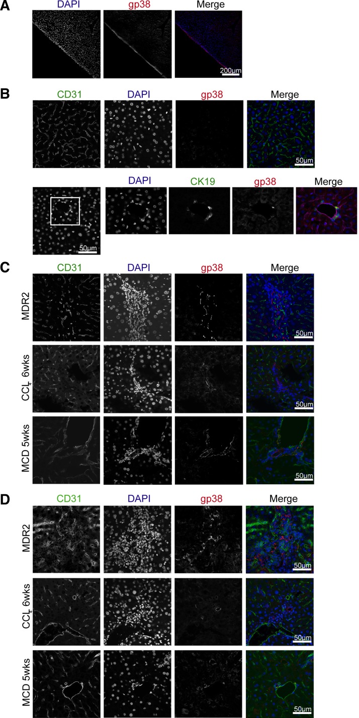
Fig. 2.
Gp38+ stromal cells localizes at cell dense structures of the injured liver. Healthy (A and B) or injured (C and D) livers were removed, preprocessed, and snap frozen. Cryosections were prepared and stained for gp38, CD31, and DAPI. Images were recorded using ×20 (B) or × 40 (A, C, and D) objectives. Scale bar = 200 μm (B) or 50 μm (A, C, and D). B, bottom: white square pinpoint area that were zoomed in. Data are representative of 2 independent experiments, n = 3 mice per group and 1 mold per organ per mouse. From each mold multiple sections were analyzed in 100- to 300-μm distances within the tissue. MCD, methionine and choline-deficient diet.

