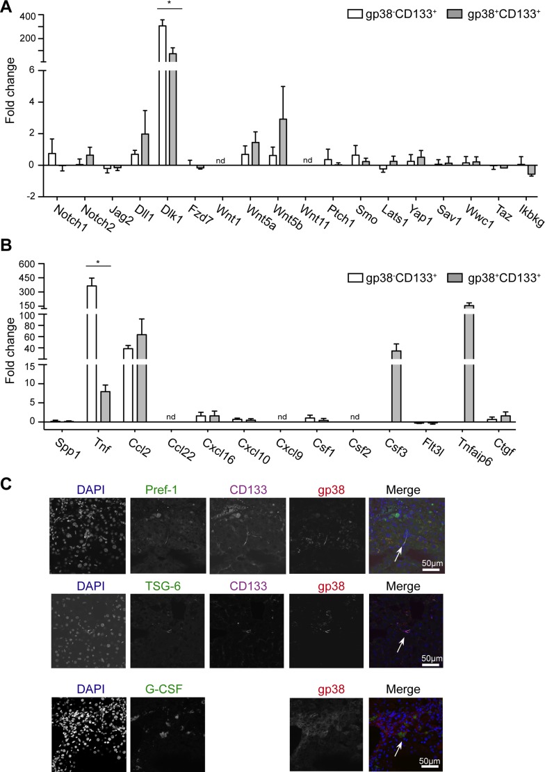
Fig. 6.
gp38+CD133+ and gp38−CD133+ cells demonstrate differences in inflammatory genes. qPCR was performed from sorted cells. Graph depicts mean fold change ± SE compared with healthy animals. Three independent sorts/subset were performed. A: genes are related to Notch, Wnt, hedgehog, and Hippo pathway. B: genes are involved in inflammation and myeloid-lymphoid cell interaction. C: cryosections from CCl4-treated liver stained for gp38, (CD133), DAPI, and either Pref-1, TSG-6, or G-SCF (×40 objectives; scale bar = 50 μm). Data are representative of 2 independent experiments, n = 3 mice/group and 1 mold/organ/mouse. *P < 0.05.

