Fig. 1.
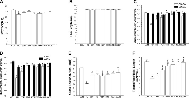
Atrophy and decreased contractile force in mouse soleus muscle after unloading and the recovery during reloading. A: 4 wk of hindlimb unloading (HU) resulted in a slight decrease of body weight, reflecting the stress effect of tail suspension. B: unloading and reloading did not produce any change in the tibial length, indicating no effect on overall growth and development. C: normalized to body weight (BW), 4 wk of HU resulted in a significant loss of soleus (SOL) muscle mass compared with that of control (CON) mice. The weight of soleus muscle recovered during reloading and reached the normal level after 15 days (15DR). EDL, extensor digitorum longus. D: same trend was shown by normalization to tibial length (TL). E: quantification of cross-sectional area (CSA) of soleus muscle confirms the atrophy after 4-wk unloading and incomplete recovery after 60 days of reloading. F: tetanic force normalized to tibial length decreased in HU soleus muscle and recovered during reloading to reach the control level after 15 days. Values are presented as means ± SE. A, B, D, and F: n = 11 in CON, n = 9 in HU, n = 9 in 3DR, n = 8 in 7DR, n = 4 in 15DR, n = 8 in 30DR, n = 7 in 45DR, and n = 7 in 60DR. C and E: n = 9 in CON, n = 4 in HU, n = 7 in 3DR, n = 6 in 7DR, n = 4 in 15DR, n = 8 in 30DR, n = 7 in 45DR, and n = 5 in 60DR. aP < 0.05 vs. CON; bP < 0.05 vs. HU; cP < 0.05 vs. 3DR. Statistical analysis was performed using 1-way ANOVA with mean comparison in Tukey's test.

