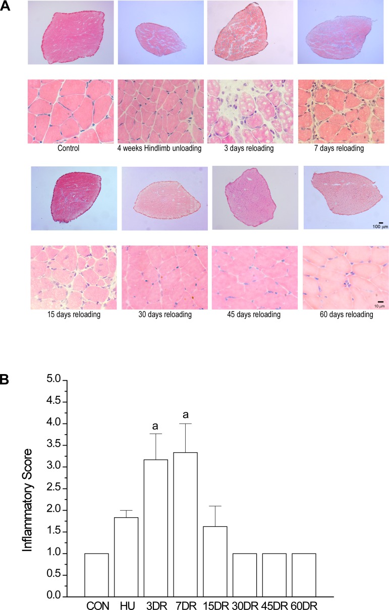
Fig. 3.
Inflammatory response in mouse soleus muscle during the early phase of reloading. A: hematoxylin and eosin staining detected infiltration of inflammatory cells in soleus muscle after 3, 7, and 15 days of reloading, indicating reloading injury of the atrophic and weakened muscle. The increased interstitial space in soleus muscle after 3 and 7 days of reloading indicates inflammatory swelling. B: using a published method (47, 68), the inflammation response to reloading injury was quantified in a blinded manner as scores of 1–4, where 1 = a few scattered inflammatory cells, 2 = clusters of inflammatory cells, 3 = diffuse infiltrate of inflammatory cells, and 4 = dense sheets of inflammatory cells, including lymphoid follicles. Values are presented as means ± SE; n = 3 in CON, n = 3 in HU, n = 3 in 3DR, n = 3 in 7DR, n = 4 in 15DR, n = 4 in 30DR, n = 4 in 45DR; and n = 3 in 60DR. aP < 0.01 vs. CON. Statistical analysis was performed using 1-way ANOVA with mean comparison in Tukey's test.

