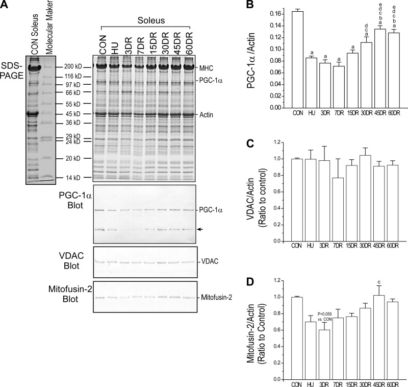
Fig. 5.
Changes of peroxisome proliferator-activated receptor γ coactivator 1α (PGC-1α), voltage-dependent anion channel (VDAC), and mitofusin-2 in mouse soleus muscles corresponding to decreased fatigue resistance after unloading and during reloading. A: Western blot using rabbit anti-PGC-1α polyclonal antibody (ab54481, Abcam) showed decreased expression of PGC-1α in mouse soleus muscle after 4 wk of unloading. B: its partial recovery during 60 days of reloading was parallel to the resistance to fatigue (a nonspecific protein band, indicated by the arrow, was also recognized by the anti-PGC-1α antibody). C: total mitochondrial content measured using a rabbit polyclonal antibody against mitochondrial VDAC was unchanged. D: however, the expression of mitochondrial fusion protein mitofusin-2 detected using a rabbit polyclonal antibody showed a similar trend to that of PGC-1α. Values are presented as means ± SE; n = 3 in CON, HU, 3DR, and 7DR; n = 4 in 15 DR; n = 4 in 30DR; n = 4 in 45DR; and n = 3 in 60DR. aP < 0.05 vs. CON; bP < 0.05 vs. HU; cP < 0.05 vs. 3DR; dP < 0.05 vs. 7DR; eP < 0.05 vs. 15DR. Statistical analysis was performed using 1-way ANOVA with mean comparison in Tukey's test.

