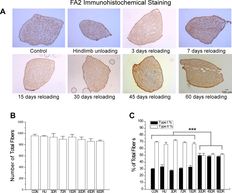
Fig. 8.
Increased number of MHC I fibers in mouse soleus muscle after 30 days of reloading. A: immunohistochemistry using mAb FA2 recognizing type I MHC determined the type I slow fiber contents of the mouse soleus muscles studied. B: total number of fibers in soleus muscle was similar in all groups, suggesting no fiber loss or hyperplasia. C: numbers of type I fibers increased, whereas type II fiber decreased after 30 days of reloading compared with the 4-wk unloading or normal control groups. This trend remained at 60 days of reloading. Values are presented as means ± SE; n = 3 in CON, n = 3 in HU, n = 3 in 3DR, n = 3 in 7DR, n = 4 in 15DR, n = 4 in 30DR, n = 4 in 45DR, and n = 3 in 60DR. ***P < 0.001 compared with the same type of fibers at different time points. Statistical analysis was performed using 1-way ANOVA with mean comparison in Tukey's test.

