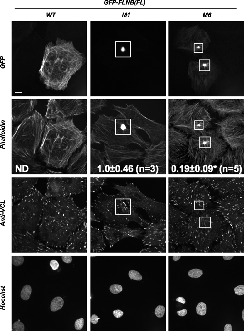
Fig. 2.
Effects of ectopic expression of GFP-FLNB on the actin cytoskeleton in Rat2 fibroblasts. Rat2 fibroblasts expressing FL GFP-FLNB WT, M1, and M6 were visualized by confocal immunofluorescence microscopy. Top to bottom: localization of GFP-FLNB, F-actin (phalloidin-Alexa Fluor 647), focal adhesions (anti-vinculin/anti-mouse IgG-Cy3), and nuclei (Hoechst dye). Boxes show location of accumulated GFP-FLNB M1 and M6. Scale bar, 10 μm. Values (means ± SE) represent F-actin accumulation index. ND, not detected. *P < 0.05 vs. M1 (by Student's t-test).

