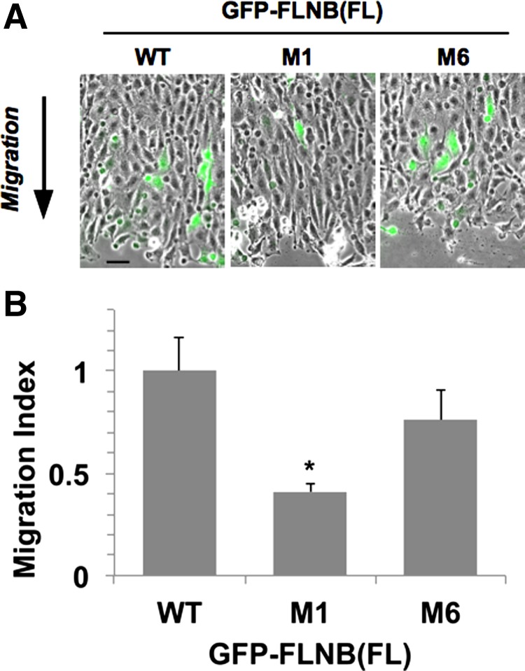
Fig. 4.
Scratch wound healing assay of Rat2 cells expressing FL GFP-FLNB WT, M1, and M6 with Venus. At 24 h after scratching, cells were fixed and stained with Hoechst dye. A: epifluorescence images of Venus signals (green) at the wound site superimposed on the corresponding phase-contrast images. Arrow indicates direction of cell migration. Scale bar, 50 μm. B: ratio of the relative population of Venus-positive cells at the wound area to that at the unscratched area (migration index). Number of Venus-positive cells in each image field was normalized against number of total cells, which was counted on Hoechst fluorescence images. Values (means ± SE from 6 image fields of 3 independent wounds) were normalized against WT. *P < 0.05 (by ANOVA with Dunnett's post hoc test).

