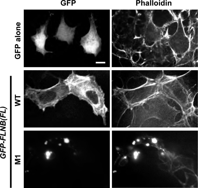
Fig. 7.
Confocal microscopy images of HEK-293 cells expressing FL FLNB WT and M1. HEK-293 cells were transiently transfected with plasmids encoding GFP alone, GFP-FLNB WT, and the M1 protein and stained with rhodamine-labeled phalloidin (1:50 dilution). Representative confocal fluorescence images of GFP and F-actin are shown. Scale bar, 10 μm.

