Figure 1.

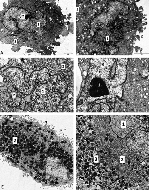
A) Electron micrograph of a dental pulp-derived stem cell from in vitro cell culture; 1, cell nucleus with predominance of euchromatin; 2, nucleolus; 3, mitochondria; 4, aggregation of glycogen granules. B) Dental pulp-derived stem cell; 1, irregular shaped nucleus with deep notches and predominance of euchromatin; 2, plasma membrane create irregular finger-like projections called fillopodia; 3, mitochondria. C) Electron micrograph of skeletal muscle-derived stem cell from in vitro cell culture; 1, pale, euchromatic nucleus; 2, dilated cisternae of rough endoplasmic reticulum. D) Detail on irregular shaped nucleus of skeletal muscle-derived stem cell, 1, large nucleolus. E) Electron micrograph of white adipose tissue-derived stem cell from in vitro cell culture; 1, pale, euchromatic nucleus; 2, residual bodies (cell organelles after degradation); 3, filopodia. F) Detail of white adipose tissue-derived stem cell from in vitro cell culture; 1, pale euchromatic nucleus; 2, cytoplasm is filled with rough endoplasmic reticulum; 3, residual bodies.
