Figure 2.

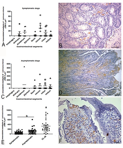
Parasite load of gastrointestinal tract (GIT) of dogs naturally infected with Leishmania (L.) infantum. A) Graph showing parasite load throughout oesophagus to rectum; in the symptomatic group, the segment of highest parasite load was the cecum, followed by the duodenum, colon, stomach, rectum, and ileum (statistical difference among groups (P≤0.001). B) Panoramic view of TGI mucosae (lower magnification) showing a presence of Leishmania amastigotes nearby muscular layer of mucosae; scale bar: 32 µm. C) Graph showing higher parasite load in duodenum and cecum in asymptomatic groups (P≤0-001). D) Higher magnification of mucosae (lamina propria) showing numerous amastigotes within macrophages brown colour highlighted by blue background colour. E) Graph showing a comparison between symptomatic, asymptomatic and CL46 dog case (P≤0.001). F) Besides a high presence of parasites, note the integrity of the epithelium lining with integrity of the intestinal glands, without ulceration or necrosis; scale bar: 16 µm. Immunohistochemistry staining by streptavidin peroxidase method.
