
Figure 9.
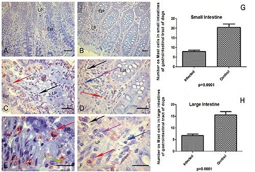
A-H) Canine tissues included in glycol methacrylate (GMA) resin of dogs naturally infected with Leishmania infantum and controls. A,B) Photomicrograph of infected dog with Leishmania (L.) infantum of ileum segment; in the lamina propria observe metachromatic mast cells (red) and epithelial cells preserved in blue background (blue toluidine); scale bars: 32 µm. C) Higher magnification showing stained metachromatic mast cells (red arrow) and plasma cells in the lamina propria (black arrow); scale bars: 16 µm. D,E,F) Observe distinct mononuclear inflammatory cells: plasma cells (black arrow), macrophages (pink arrow), lymphocytes (blue arrow) and mast cells (red arrow); rare neutrophils could be seen in lamina propria (green arrows); scale bars: D) 16 µm; E,F) 8 µm. G) Graph showing a lower number of mast cells in small intestine (duodenum, jejunum and ileum) of all infected dogs (P≤0.0001). H) Graph showing a lower number of mast cells in large intestine (caecum, colon and rectum) of all infected dogs groups (P≤0.0001).