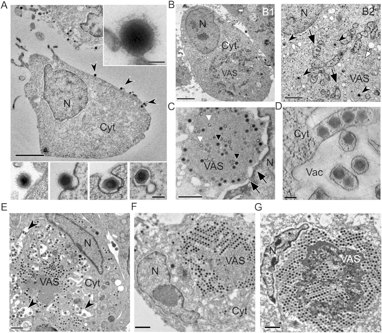
Figure 1. Electron microscopy of high-pressure freezing, freeze substitution of Singapore Grouper Iridovirus (SGIV) infected grouper embryonic cells (GECs) reveals the SGIV life cycle.
(A) SGIV attaches onto a GEC at 30 min post-infection (arrowheads); scale bar = 1μm. The outermost layer (top inset) around the capsid may initiate the entry process, which takes place through endocytosis (bottom inset, 1 hpi); scale bar = 200 nm. N = nucleus, Cyt = cytoplasm. (B) At 1 hpi, the viral assembly site (VAS) starts to form (B1, scale bar = 1μm). Many SGIV particles are still contained in coated vesicles, which is likely to represent SGIV particles in transit (B2, arrowheads, scale bar = 200 nm). Already, some viral intermediates such as membrane filaments can be observed (B2, white arrow). At this time point, mitochondria start to accumulate at the VAS boundary. Some mitochondria appear to have lost their internal structure (B2, black arrows). B2 is an enlarged area of B1. (C) At 4–12 hpi, the VAS and nucleus are now interconnected via nuclear pores (arrows), and many partially filled capsids (white triangle) and fully filled capsids (black triangle) are present; scale bar = 1 μm. (D,E) At 8 hpi, the VAS has expanded dramatically causing the nucleus to deform, and mature virions have accumulated at the edge of the VAS where large vacuoles have formed. Some mature viruses appear to bud into the newly-formed vacuoles containing membrane tubules (E, arrowheads; scale bar = 1 μm). The picture D is 220 nm thick tomographic slice showing membrane tubules with a row of mature virions; scale bar = 200 nm. (F,G) At 48 hpi, the late stage of infection the VAS markedly enlarges and occupies majority of the cell. Mature virions densely accumulate and become packed in the form of paracrystalline arrays inside the cell. The nucleus is pushed to the margin of the cell and now has a highly deformed appearance. The release of mature virions takes place through budding as enveloped virions or through cell lysis as unenveloped virions; scale bars = 1 μm.

