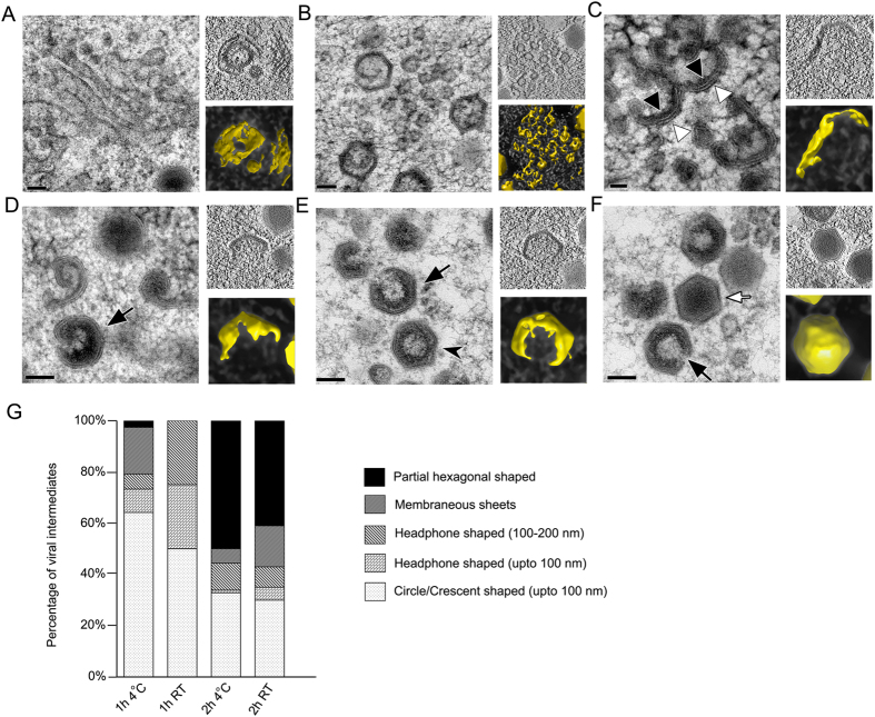
Figure 2. Morphogenesis of SGIV capsids in the VAS.
(A–F) Main panel, electron micrograph depicting different viral intermediates during capsid assembly; Top right panel, insets of the central slice through a tomogram; Bottom right panel, isosurface rendition of the corresponding viral intermediates (yellow). (A,B) Membranous vesicles, tubules, and sheets, scale bar = 100 nm. (C) Crescent-shaped structures with two shells; scale bar = 50 nm. (D–F) Electron dense materials accumulate on the inner layer as the capsid is being filled. As the packing process continues, the two shell crescent-shaped structure curves to form icosahedral capsids with an opening (black arrow). Mature virions (white arrow) with a dense core are eventually formed. No opening was observed on the fully formed mature particles; scale bars = 100 nm. (G) The distribution of capsid intermediates observed in infected GECs maintained at 4 °C and 27 °C, at 1 or 2 hpi. Images of the representative intermediates are shown in Fig S1.

